Retina Expert Witness
A retina expert witness evaluates whether retinal disease was diagnosed, monitored, and treated within the standard of care, and whether an alleged departure actually caused the claimed vision loss. Marc H. Shomer, MD, PhD, QME, a board-certified ophthalmologist, reviews retinal claims for plaintiff and defense counsel nationwide, drawing on more than 13 years of medical-legal consulting experience.
Retinal cases reward careful record work because the retina, uniquely among injured tissues, is photographed and scanned in ordinary clinical practice. The images in the chart often answer the timing questions that the parties dispute.
The Medical-Legal Question in Retinal Cases
Most retinal litigation reduces to two questions: was the disease detected and treated when a reasonable ophthalmologist would have detected and treated it, and if not, how much of the ultimate visual outcome is attributable to the delay rather than to the underlying disease? The retina does not regenerate. Once photoreceptors are lost, to ischemia, to detachment, to untreated leakage, the deficit is generally permanent, which is why timing dominates both liability and damages analysis.
Matters Where a Retina Expert Is Retained
- Diabetic retinopathy: alleged failures of screening, monitoring intervals, or timely laser and anti-VEGF (anti-vascular endothelial growth factor) treatment in patients with progressing disease.
- Retinal vascular occlusions, central and branch retinal artery or vein occlusions, including whether an artery occlusion was recognized as a stroke-equivalent event requiring urgent systemic workup.
- Macular disorders, age-related macular degeneration and related conditions, addressed in depth on the macular disease page.
- Retinal detachment, the most common delayed-diagnosis theme, covered on the dedicated retinal detachment expert witness page.
- Trauma and toxicity, retinal injury after blunt or penetrating trauma, and drug-related maculopathy.
Why Retinal Imaging Is Powerful Evidence
Two technologies give retinal cases an unusual degree of objectivity. Optical coherence tomography (OCT) is a non-invasive scan that produces cross-sectional images of the retina at near-microscopic resolution; it documents fluid, atrophy, and structural damage in a form that does not depend on the patient's effort or report. Fundus photography records the visible surface of the retina, so hemorrhages, exudates, and detachments can be compared across dates. When serial images exist, an expert can often reconstruct when disease appeared, how fast it progressed, and what an examiner on a given date could reasonably have seen. The attorney-resources page on OCT as evidence explains how these studies are read.
Causation and Standard-of-Care Considerations
Association is not causation. A patient whose diabetic macular edema worsened between visits may have deteriorated because of a monitoring lapse: or because the disease was aggressive, systemic control was poor, or treatment was declined. An honest analysis considers the natural history of the specific condition, the visual acuity and imaging at each decision point, and what earlier intervention would more likely than not have changed. Some occlusions cause profound, immediate, untreatable vision loss; attributing that outcome to a later clinical decision misstates the medicine. These questions overlap with the general frameworks discussed on the causation and standard of care pages.
What an Honest Evaluation Can and Cannot Determine
Where imaging is dense, opinions can be anchored to dated, objective structure. Where the record is sparse, a single undilated exam, no photographs, gaps in follow-up, the expert must say plainly what cannot be reconstructed. Retinal prognosis also carries genuine uncertainty: two eyes with similar scans can follow different courses, and a credible report expresses that range rather than false precision.
What Attorneys Should Provide
Complete ophthalmology and optometry records including the actual OCT scans and fundus photographs (not just the reports), primary care and endocrinology records for diabetic patients, operative reports for any retinal surgery, and both pre- and post-event records so the baseline is documented. The records checklist covers the details. Dr. Shomer provides record review, chronologies, written reports, and deposition and trial testimony for both plaintiff and defense matters.
Frequently Asked Questions
What does a retina expert witness do?
A retina expert witness reviews the clinical records and retinal imaging in a case involving retinal disease or injury, then offers opinions on whether care met the standard of care and whether any departure caused the claimed vision loss. The work typically includes record review, a written report, and deposition or trial testimony.
Why is OCT imaging so important in retinal litigation?
Optical coherence tomography (OCT) produces objective, dated, cross-sectional images of retinal structure that do not depend on patient effort or recollection. Serial OCT scans can show when damage appeared and how it progressed, which often anchors the timing questions at the center of delayed-diagnosis claims.
Does delayed treatment of retinal disease always cause worse vision?
No. Some retinal conditions cause immediate, largely untreatable vision loss regardless of response time, while others are highly time-sensitive. A credible causation opinion compares the documented state of the retina at each decision point with the natural history of the specific disease, rather than assuming any delay changed the outcome.
Does Dr. Shomer review retinal cases for both plaintiffs and defendants?
Yes. Dr. Shomer has consulted on both plaintiff and defense matters for more than 13 years, and the analysis is the same either way: what the records and imaging objectively show, measured against the standard of care and mainstream ophthalmology.
Educational information only. This page provides general information for attorneys and other medical-legal professionals. It is not medical or legal advice, does not address any particular case, and does not create a physician-patient, attorney-client, or expert-client relationship. Opinions in any matter are formed only after review of the specific records, examination findings, and applicable literature. Past engagements do not guarantee any result.
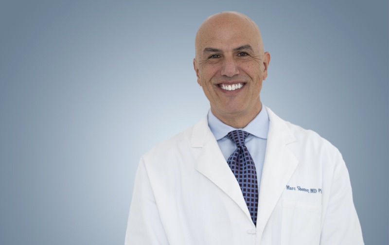
Board-certified ophthalmologist; Adjunct Associate Professor of Ophthalmology, Keck School of Medicine of USC; active California Qualified Medical Evaluator. Full biography · Curriculum vitae
Published August 2, 2026 · Last substantive review August 2, 2026 · Medical reviewer: Marc H. Shomer, MD, PhD, QME
Request a Conflict Check and Case Review
Submit the matter for conflict screening. Please do not send medical records or protected health information until conflicts are cleared and secure transfer instructions are provided.