Neuro-Ophthalmology Expert Witness
A neuro-ophthalmology expert witness addresses cases where the visual problem originates not in the eye itself but in the optic nerves, visual pathways, or the nerves controlling eye movement: territory where ophthalmology, neurology, and neurosurgery overlap and where diagnoses are missed at the seams between specialties. Marc H. Shomer, MD, PhD, QME, a board-certified ophthalmologist with a PhD in neuroscience from UCLA, reviews these matters for plaintiff and defense counsel.
Neuro-ophthalmic cases are high-stakes because the eye finding is often the visible marker of intracranial disease: the question is rarely just about vision, but about what the eye exam should have revealed about the brain.
The Medical-Legal Question
These cases typically ask whether an eye or systemic finding, a swollen optic nerve, a new double vision, a field cut, was recognized as the sign of underlying neurologic disease it was, and whether the required workup and referral happened in time. Because the underlying conditions include tumors, aneurysms, elevated intracranial pressure, and stroke, a missed neuro-ophthalmic sign can translate into injury far beyond the eye.
Recurring Case Types
- Papilledema and missed intracranial pathology. Papilledema is swelling of both optic nerve heads from elevated pressure inside the skull. It is visible on a routine dilated exam and is an examination finding that demands urgent neuroimaging and workup. Claims arise when disc swelling was not detected, was documented but not pursued, or when headaches were managed for months without fundus examination.
- Cranial nerve palsies and diplopia after trauma. The third, fourth, and sixth cranial nerves steer the eyes; injury to any of them causes misalignment and double vision (diplopia). After head trauma, the questions include which nerve was injured, whether the pattern fits the trauma, whether an underlying compressive lesion was excluded, a third nerve palsy with pupil involvement raises aneurysm until proven otherwise, and the prognosis for recovery or strabismus surgery.
- Visual pathway injuries. Damage behind the eyes, chiasm, optic radiations, visual cortex, produces characteristic field defects, such as loss of the same half of the field in both eyes (homonymous hemianopia). Mapping the defect localizes the lesion, which bears directly on causation in trauma and stroke cases.
- Giant cell arteritis. This inflammatory disease of medium and large arteries in patients generally over 50 can cause sudden, permanent blindness, and the second eye may follow the first within days if steroids are not started promptly. It is a true ophthalmic emergency, and litigation centers on whether classic symptoms, new headache, scalp tenderness, jaw pain with chewing, transient visual loss, prompted timely testing and treatment.
Distinguishing Ocular From Neurologic Visual Loss
A core skill in this area is localization: deciding whether visual loss arises in the eye, the optic nerve, or the brain. The tools are the pupil exam, a relative afferent pupillary defect (RAPD) indicates asymmetric optic nerve dysfunction, color vision testing, formal visual fields whose patterns localize the lesion, OCT (optical coherence tomography) of the nerve fiber layer, and neuroimaging. The same toolkit identifies non-organic vision loss, where reported deficits are inconsistent with objective structure and physiology. Field interpretation is covered in the primer on understanding visual fields, and optic nerve injury specifically on the optic nerve injury page.
Causation Considerations and Honest Limits
Temporal association is seductive in this area, visual symptoms after a car accident, a palsy after a procedure, but microvascular nerve palsies occur spontaneously in patients with diabetes and hypertension, and many field defects predate the event that brought them to attention. The analysis weighs the lesion's localization against the alleged mechanism, as outlined on the causation page. Prognostic statements about pathway injuries also carry real uncertainty, particularly early after injury, and a responsible report says so.
What Attorneys Should Provide
Neuroimaging (the actual MRI and CT studies, not just reports), emergency department and neurology records, all ophthalmic records with visual fields and OCT, laboratory results (including inflammatory markers in suspected giant cell arteritis), and prior records establishing baseline vision and alignment. Dr. Shomer provides record review, written reports, and deposition and trial testimony nationwide for both sides of the bar.
Frequently Asked Questions
What is a neuro-ophthalmology expert witness?
An expert who evaluates visual problems originating in the optic nerves, visual pathways, or eye-movement nerves rather than the eye itself: such as papilledema, cranial nerve palsies, hemianopic field loss, and giant cell arteritis. These cases sit at the boundary between ophthalmology and neurology, where diagnostic responsibility is often disputed.
Why is papilledema such a frequent malpractice theme?
Papilledema, optic nerve swelling from elevated intracranial pressure, is visible on an ordinary dilated eye exam and signals potentially serious intracranial disease requiring urgent imaging. When headaches or visual complaints are managed for months without a fundus examination, or documented disc swelling is not pursued, delayed diagnosis of tumors or raised intracranial pressure can result.
Is giant cell arteritis really an eye emergency?
Yes. Giant cell arteritis can cause sudden, irreversible blindness, and without prompt corticosteroid treatment the second eye can be lost within days of the first. Because prompt treatment protects the fellow eye, timing of diagnosis and steroid initiation is usually the central standard-of-care and causation question in these cases.
Can testing distinguish neurologic vision loss from an eye problem?
Usually. Pupillary testing, color vision, the pattern of visual field loss, OCT of the optic nerve, and neuroimaging together localize the problem to the eye, the optic nerve, or the brain. The same objective toolkit identifies claimed deficits that are inconsistent with the underlying anatomy and physiology.
Educational information only. This page provides general information for attorneys and other medical-legal professionals. It is not medical or legal advice, does not address any particular case, and does not create a physician-patient, attorney-client, or expert-client relationship. Opinions in any matter are formed only after review of the specific records, examination findings, and applicable literature. Past engagements do not guarantee any result.
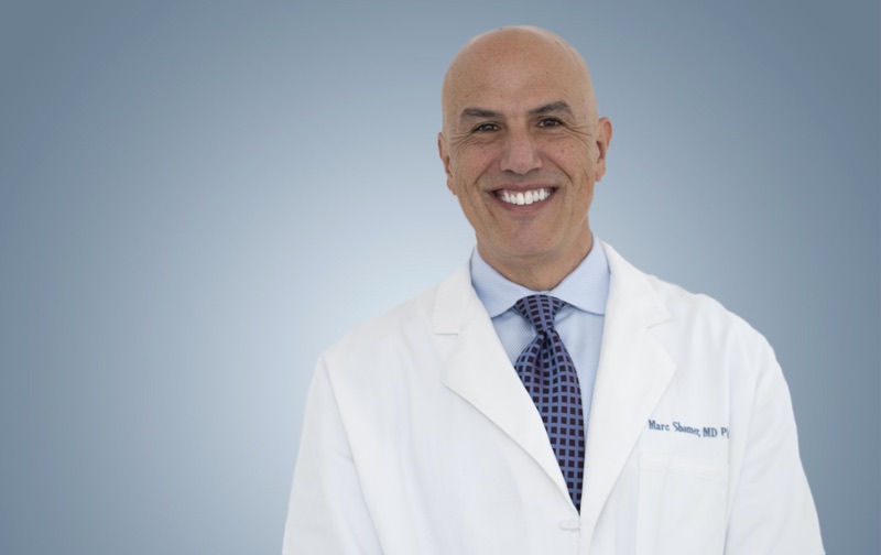
Board-certified ophthalmologist; Adjunct Associate Professor of Ophthalmology, Keck School of Medicine of USC; active California Qualified Medical Evaluator. Full biography · Curriculum vitae
Published August 2, 2026 · Last substantive review August 2, 2026 · Medical reviewer: Marc H. Shomer, MD, PhD, QME
Request a Conflict Check and Case Review
Submit the matter for conflict screening. Please do not send medical records or protected health information until conflicts are cleared and secure transfer instructions are provided.