OCT as Objective Evidence in Eye Injury Litigation
Optical coherence tomography (OCT) is a non-invasive scan that produces cross-sectional images of the retina at near-microscopic resolution, and it is among the most powerful objective evidence available in eye injury litigation because it documents anatomy directly, independent of what the examinee reports. Where acuity and visual field tests record what a person says or does, OCT records what the tissue is.
What OCT Is
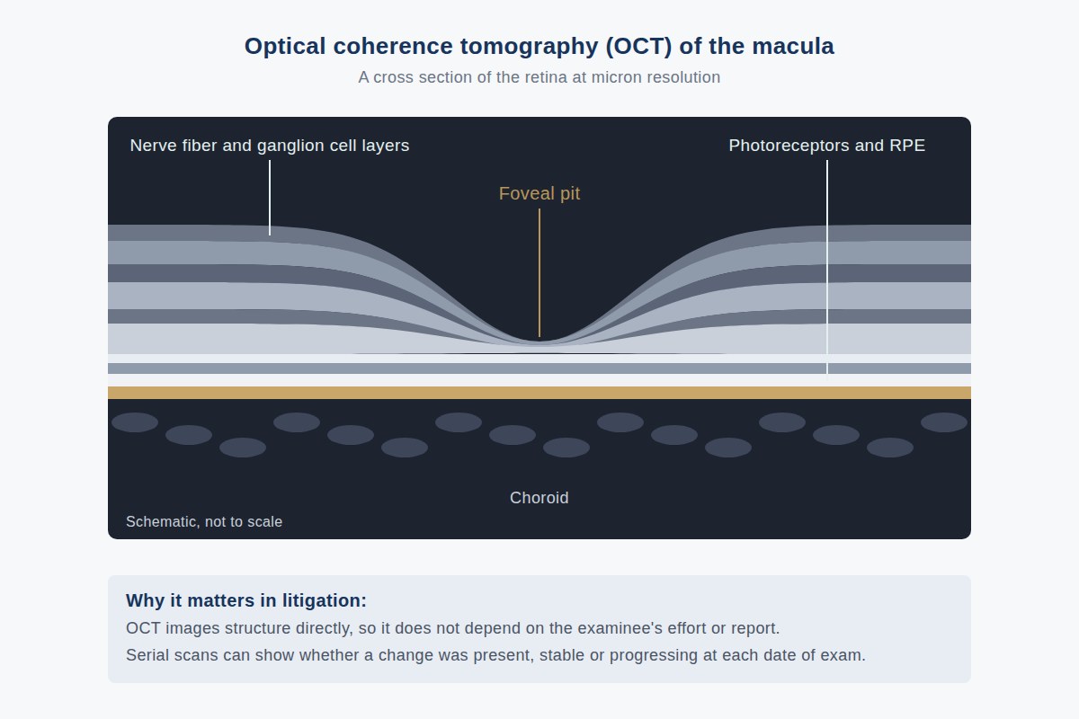
OCT works on a principle analogous to ultrasound, using reflected light instead of sound. The instrument sweeps a light beam across the back of the eye and measures reflections from each tissue depth, assembling cross-sectional images, in effect, optical biopsies, with resolution measured in microns. The scan takes seconds, requires nothing more of the examinee than looking at a target, and is performed routinely in ophthalmic practice, which means most litigants with eye complaints already have OCT data in their records.
What OCT Shows
The retina is a layered structure, and OCT renders those layers individually: the photoreceptors that capture light, the intermediate processing layers, and the nerve fiber layer whose axons form the optic nerve. Clinically important measurements include:
- Macular anatomy, the central retina responsible for reading vision. OCT shows swelling (edema), holes, scar tissue, fluid under the retina, and degeneration with a clarity no ophthalmoscope achieves. These findings anchor disputes about macular disease and surgical complications.
- Retinal nerve fiber layer (RNFL) thickness, measured around the optic nerve and compared to normative data. Thinning marks permanent axonal loss from glaucoma, optic nerve injury, or other optic neuropathy.
- Serial change, successive scans on the same instrument can be registered and compared, quantifying progression or stability over time.
Why It Is Powerful Evidence
OCT has three properties that make it valuable in litigation. It is objective: the examinee cannot will a retinal layer to look thinner or thicker, so the scan is immune to the effort and secondary-gain problems that complicate acuity and field testing. It is quantitative: thicknesses are measured in microns against normative databases, allowing precise comparisons between eyes, between visits, and against normal. And it is ubiquitous: because OCT is standard of care for many conditions, contemporaneous scans often exist from before, at, and after the events in dispute, evidence created for clinical purposes, with no litigation in view.
The Timing Value: Structural Change Dates Injury
Structural findings evolve on characteristic timescales, and this gives OCT a chronological dimension. After an optic nerve insult, RNFL thinning develops over roughly weeks to months as injured axons degenerate: so a scan showing established, stable thinning shortly after an alleged injury suggests the loss predates it, while normal thickness immediately after trauma followed by progressive thinning on serial scans is consistent with the injury as the cause. Similar logic applies to macular findings: chronic degenerative changes look different from acute ones. An OCT taken years before an incident can establish a pristine baseline; one taken the week after can show whether the claimed damage was already old. This dating function, structure as a timestamp, is frequently the decisive contribution of OCT to causation analysis.
Limitations: What OCT Cannot Do
An honest evaluation acknowledges the technique's boundaries:
- Artifacts. Blinks, eye movement, media opacity (dense cataract, vitreous debris), and poor signal strength degrade scans. A low-quality scan can mimic or mask pathology, which is why signal-strength metrics on the printout matter.
- Segmentation errors. The instrument's software draws the boundaries between retinal layers automatically, and it can draw them wrong, particularly in distorted or diseased retinas. Erroneous segmentation produces erroneous thickness numbers. Checking the machine's line placement against the raw image requires the native scan data, not a summary printout.
- Structure is not function. OCT measures anatomy, not vision. A structurally normal scan does not by itself prove normal sight (some pathology is invisible to OCT, and the visual cortex lies beyond its reach), and measurable thinning does not by itself establish disabling loss. Function is measured by acuity, fields, and electrophysiology; the strongest analyses correlate the two rather than substituting one for the other.
- Floor effects. In long-standing severe atrophy, RNFL measurements bottom out and can no longer track further worsening.
Key point: OCT is objective, quantitative, and often pre-existing in the clinical record, but its evidentiary value depends on scan quality and correct segmentation, which can only be verified from native data files. Printouts of OCT studies discard the information needed to audit them.
What Attorneys Should Provide
Request every OCT study ever performed on the claimant, from every provider, in native format, together with the visual function testing from the same period. Serial scans from the same instrument are especially valuable because the software can register them for direct comparison. Guidance on obtaining native imaging appears in What Records to Provide.
Frequently Asked Questions
What is OCT in simple terms?
Optical coherence tomography is a quick, non-invasive scan that uses reflected light to produce cross-sectional images of the retina at near-microscopic resolution, an optical biopsy of the back of the eye. It objectively documents retinal structure regardless of what the examinee reports.
Why is OCT considered objective evidence?
Because it measures tissue anatomy directly, an examinee cannot influence OCT results through effort or reporting, unlike acuity and visual field tests, which depend on responses. Thicknesses are quantified in microns against normative databases, permitting precise comparison between visits and against normal values.
Can OCT tell when an eye injury occurred?
Often it can bracket the timing. Structural changes such as retinal nerve fiber layer thinning develop over characteristic intervals after injury, so the presence, absence, and progression of findings on dated scans can indicate whether damage predated an incident or developed after it. Serial scans strengthen this analysis considerably.
What are the limitations of OCT evidence?
OCT is subject to artifacts from movement, blinking, and media opacity; its automated layer segmentation can err, producing wrong thickness values; and it measures structure, not visual function. Sound opinions verify scan quality from native data and correlate OCT findings with functional testing rather than relying on either alone.
Educational information only. This page provides general information for attorneys and other medical-legal professionals. It is not medical or legal advice, does not address any particular case, and does not create a physician-patient, attorney-client, or expert-client relationship. Opinions in any matter are formed only after review of the specific records, examination findings, and applicable literature. Past engagements do not guarantee any result.

Board-certified ophthalmologist; Adjunct Associate Professor of Ophthalmology, Keck School of Medicine of USC; active California Qualified Medical Evaluator. Full biography · Curriculum vitae
Published August 2, 2026 · Last substantive review August 2, 2026 · Medical reviewer: Marc H. Shomer, MD, PhD, QME
Request a Conflict Check and Case Review
Submit the matter for conflict screening. Please do not send medical records or protected health information until conflicts are cleared and secure transfer instructions are provided.