Traumatic Brain Injury Vision Expert Witness
A traumatic brain injury vision expert witness evaluates the visual complaints that follow a head injury: blurred or double vision, difficulty reading, light sensitivity and loss of side vision, often in a patient whose eyes look normal on a routine examination. Marc H. Shomer, MD, PhD, QME, a board-certified ophthalmologist with a PhD in neuroscience from UCLA, examines these patients and reviews these matters for plaintiff and defense counsel and, in California, as a Qualified Medical Evaluator.
These are among the most contested vision claims in litigation, for an understandable reason. The symptoms are real and common after head injury, yet many of them cannot be seen by looking at the eye, and the standard measure of vision, the letter chart, is usually normal. The work of the expert is to establish which complaints have objective support, which are consistent with the documented injury, and which have another explanation.
What the research shows
A systematic review and meta-analysis of 22 studies of patients with traumatic brain injury and no eye injury reported pooled prevalence estimates of 42.8 percent for accommodative dysfunction (impaired focusing), 36.3 percent for convergence insufficiency (difficulty turning the eyes inward together for near work) and 18.2 percent for visual field loss. Loss of acuity to the level of legal blindness was essentially absent. Visual field loss was far more common after moderate to severe injury (39.8 percent) than after mild injury (6.6 percent).
Two points follow. A normal 20/20 acuity does not rule out a visual consequence of head injury. And the type of deficit should fit the severity of the injury: a dense field defect after a mild concussion with normal imaging calls for careful scrutiny, while reading fatigue from a focusing or convergence deficit is entirely plausible.
How head injury affects vision
- Focusing and eye teaming. Accommodative and vergence deficits produce near blur, eyestrain, words that move or double on the page, and headaches with reading. These were the most common findings in a review of 160 patients with traumatic brain injury and visual symptoms.
- The nerves that move the eyes. Injury to the third, fourth or sixth cranial nerve misaligns the eyes and causes double vision. The misalignment can be measured in prism diopters in each direction of gaze, and the pattern identifies the nerve.
- The optic nerve. Traumatic optic neuropathy causes loss of acuity, color vision and visual field in one eye, with an abnormal pupil response. Weeks later the nerve becomes pale and OCT (optical coherence tomography) shows thinning. See Optic Nerve Injury.
- The visual pathways in the brain. Damage behind the eyes produces matching field loss in both eyes, typically on the same side. The defect should correspond to the location of injury on imaging. See Visual Field Loss.
- Light sensitivity and visual discomfort. Common after concussion and largely subjective. They are documented by history and by excluding ocular causes such as dry eye and inflammation.
- The eye itself. The force that injured the brain may have injured the eye. A dilated examination is needed to exclude retinal tears, lens injury and damage to the drainage angle. See Ocular Trauma.
Objective evidence and its limits
Several findings do not depend on what the patient reports. A relative afferent pupillary defect indicates damage to one optic nerve. OCT measures the nerve fiber and ganglion cell layers, and structural loss after optic nerve injury persists permanently. Eye misalignment can be observed and measured. Visual field tests carry reliability indices, and genuine neurologic field loss follows anatomical rules, such as respecting the vertical midline.
The limits matter just as much. Injury to the visual pathways deep in the brain may leave the OCT normal. Focusing ability declines naturally with age, so a reduced measurement in a patient over forty may be presbyopia. Convergence testing depends on effort. An honest report states which findings are objective, which are effort-dependent, and how the two fit together. The approach to inconsistent findings is described in Non-Organic Visual Loss: What the Testing Shows.
Normal acuity is the beginning of the evaluation. A complete examination after head injury includes pupils, color vision, eye alignment and movement at distance and near, focusing and convergence measurements, formal visual fields, a dilated fundus examination and OCT of the optic nerve and macula. A file that contains only a letter-chart acuity cannot answer the question either side is asking.
Causation
The comparison is always against the patient’s vision before the injury. Prior optometry and ophthalmology records establish the baseline prescription and any pre-existing eye turn, lazy eye or field defect. A long-standing tendency of the eyes to drift can decompensate after a head injury, and can also decompensate with age and fatigue. Medications prescribed after the injury can impair focusing. The timing of the first complaint matters: symptoms recorded in the early emergency, neurology or therapy notes carry more weight than symptoms that first appear many months later.
Treatment questions
For traumatic optic neuropathy, the International Optic Nerve Trauma Study found no clear benefit from corticosteroids or optic canal surgery and concluded that neither should be considered the standard of care. An allegation that vision was lost because steroids were not given therefore faces a real evidentiary obstacle. For focusing and convergence deficits, treatment may include glasses, prisms and therapy, and the reasonableness and duration of that care are often part of the damages dispute.
The scope of Dr. Shomer’s opinion
Dr. Shomer confines his opinion to the visual system: what the examination and testing show, whether the findings are consistent with the documented injury, and what visual impairment results. He relies on the neurologic and imaging record for the diagnosis of the brain injury itself, and he does not offer opinions on cognition, balance or psychological injury, which belong to other specialties. For permanent impairment, visual acuity, visual field and double vision are rated as described at Visual Disability Evaluation. Related neurologic eye findings are covered at Neuro-Ophthalmology.
Frequently Asked Questions
Can someone have 20/20 vision and still have a visual injury from a head injury?
Yes. A meta-analysis of patients with traumatic brain injury found that loss of acuity to the level of legal blindness was essentially absent, while focusing problems, convergence insufficiency and visual field loss were common. The letter chart does not measure those functions.
What objective tests support a vision claim after head injury?
The pupil examination, OCT measurement of the optic nerve fiber and ganglion cell layers, measurement of eye misalignment, and visual field testing with reliability indices. Each has limits: for example, injury to the visual pathways deep in the brain may leave the OCT normal.
Is failure to give steroids for traumatic optic neuropathy a breach of the standard of care?
The International Optic Nerve Trauma Study found no clear benefit from corticosteroids or optic canal surgery and concluded that neither should be considered the standard of care, and that it is reasonable to decide whether to treat on an individual basis.
How common is visual field loss after a mild head injury?
It is uncommon. In a meta-analysis, visual field loss was found in 6.6 percent of patients with mild traumatic brain injury compared with 39.8 percent of those with moderate to severe injury. A significant field defect after a mild injury calls for correlation with imaging and with objective eye findings.
Authorities Cited
Primary sources referenced on this page. Links open the source on the publisher’s or agency’s site.
Educational information only. This page provides general information for attorneys and other medical-legal professionals. It is not medical or legal advice, does not address any particular case, and does not create a physician-patient, attorney-client, or expert-client relationship. Opinions in any matter are formed only after review of the specific records, examination findings, and applicable literature. Past engagements do not guarantee any result.
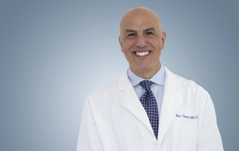
Board-certified ophthalmologist; Adjunct Associate Professor of Ophthalmology, Keck School of Medicine of USC; active California Qualified Medical Evaluator. Full biography · Curriculum vitae
Published October 5, 2026 · Last substantive review October 5, 2026 · Medical reviewer: Marc H. Shomer, MD, PhD, QME
Request a Conflict Check and Case Review
Submit the matter for conflict screening. Please do not send medical records or protected health information until conflicts are cleared and secure transfer instructions are provided.