Blunt Eye Trauma
Blunt eye trauma, from a fist, ball, airbag, or fall, compresses the globe without breaching its wall, and the resulting damage can involve nearly every structure of the eye at once. The medical-legal challenge is twofold: some consequences appear immediately and heal, while others, most notably angle-recession glaucoma, may not declare themselves for years. Sound causation and damages opinions therefore depend on longitudinal follow-up, not just the emergency-room snapshot.
Dr. Marc H. Shomer, a board-certified ophthalmologist and California QME with more than 13 years of medical-legal experience, evaluates blunt trauma claims for plaintiff and defense counsel.
How Blunt Force Injures the Eye
A blunt impact briefly compresses the globe front-to-back and expands it side-to-side. That mechanical distortion shears the delicate structures inside: the iris root, the fine fibers suspending the lens, the drainage angle, the retina, and the choroid beneath it. One blow can therefore produce a constellation of findings, and the documented pattern should be internally consistent with a compressive mechanism, a consistency check that is part of any careful review, as discussed on the ocular trauma expert witness hub page.
The Characteristic Sequelae
- Hyphema, blood in the anterior chamber, the space between cornea and iris. Usually resolves, but signals significant force and marks the eye for angle injury; rebleeding and pressure spikes are the acute management issues.
- Angle recession, a tear within the eye's internal drainage angle, visible on gonioscopy (examination of the angle with a mirrored contact lens). It matters because extensive recession predisposes to late-onset glaucoma, sometimes years or decades after the injury.
- Iridodialysis, separation of the iris root from its attachment, producing an irregular pupil and often glare complaints.
- Traumatic cataract and lens dislocation, clouding of the natural lens, or its displacement when the suspending fibers tear; either may require surgery whose complexity exceeds routine cataract surgery.
- Commotio retinae, retinal whitening from the shock wave. Often transient, but foveal involvement can leave permanent central-vision deficit.
- Choroidal rupture: a tear in the vascular layer beneath the retina; when it crosses near the macula, permanent central vision loss can result, and late complications can arise at the rupture site.
- Orbital fracture, breakage of the thin bones around the eye, with possible double vision from muscle entrapment or a sunken appearance; associated globe injury must always be excluded.
Records and Examination Components
The acute record should contain presenting visual acuity, pressure measurements, and a dilated fundus examination; the follow-up record should contain gonioscopy once the eye quiets, angle recession cannot be documented without it, plus serial pressures, OCT (optical coherence tomography, cross-sectional retinal imaging) of the macula where central vision is at issue, and photographs. In an in-person evaluation, Dr. Shomer performs these examinations directly. Pre-incident records remain essential: cataracts and glaucoma also occur without trauma, and the pre-injury baseline is the anchor for attribution.
Causation and the Long Tail of Blunt Trauma
The signature causation problem here is latency. A claimant may develop glaucoma five, ten, or twenty years after a documented hyphema; whether that glaucoma is traumatic turns on the documented angle findings, the asymmetry between the two eyes, and the exclusion of ordinary open-angle glaucoma, which is common in the general population. Conversely, a cataract appearing in a sixty-year-old three years after a minor contusion may owe more to age than to injury. This apportionment reasoning is the daily work of workers' compensation evaluation, see workers' compensation eye injury, and of civil causation analysis alike.
Key point: Blunt trauma's most serious consequences can be delayed by years. Longitudinal records, especially gonioscopy findings and serial pressures, often decide whether a late condition is traumatic or coincidental.
What an Evaluation Can and Cannot Determine
An examination years after injury can document current structure and function with precision, and permanent findings such as angle recession or a choroidal rupture scar are durable evidence of past trauma. What no examination can do is retroactively establish findings that were never recorded: if gonioscopy was never performed, the angle status at the time of injury is simply unknown, and the opinion must carry that uncertainty honestly.
What Attorneys Should Provide
Emergency records, all subsequent ophthalmology and optometry records including gonioscopy notes and pressure logs, imaging and photographs in native form, pre-incident eye records going back as far as available, and a description of the mechanism (object, distance, protective eyewear). The attorney checklist for eye injury cases provides a fuller list.
Frequently Asked Questions
What is angle recession and why does it matter in litigation?
Angle recession is a tear within the eye's internal drainage structure caused by blunt trauma, detectable on gonioscopy. It matters because extensive recession predisposes to glaucoma that may not appear for years or decades, making it a frequent link, or claimed link, between an old injury and later vision loss.
Can glaucoma diagnosed years after an eye injury be caused by that injury?
It can be. Traumatic angle damage can produce glaucoma long after the original injury. Attribution depends on documented angle recession, asymmetry between the injured and uninjured eye, and exclusion of ordinary open-angle glaucoma, which is common without any trauma. The follow-up record usually decides the question.
Is a traumatic cataract different from an age-related cataract?
Blunt trauma can cloud the lens in patterns ophthalmologists recognize as traumatic, and injury can also displace the lens by tearing its supporting fibers. In older claimants the analysis must separate injury effect from age-related change, using the pre-incident records and the comparison with the fellow eye.
Educational information only. This page provides general information for attorneys and other medical-legal professionals. It is not medical or legal advice, does not address any particular case, and does not create a physician-patient, attorney-client, or expert-client relationship. Opinions in any matter are formed only after review of the specific records, examination findings, and applicable literature. Past engagements do not guarantee any result.
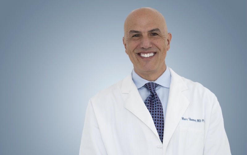
Board-certified ophthalmologist; Adjunct Associate Professor of Ophthalmology, Keck School of Medicine of USC; active California Qualified Medical Evaluator. Full biography · Curriculum vitae
Published August 2, 2026 · Last substantive review August 2, 2026 · Medical reviewer: Marc H. Shomer, MD, PhD, QME
Request a Conflict Check and Case Review
Submit the matter for conflict screening. Please do not send medical records or protected health information until conflicts are cleared and secure transfer instructions are provided.