Optic Nerve Injury Expert Witness
An optic nerve injury expert evaluates whether damage to the optic nerve, the structure that carries all visual information from eye to brain, was caused by the alleged trauma, surgery, or disease process, and what the objective findings say about when the injury occurred. Marc H. Shomer, MD, PhD, QME, a board-certified ophthalmologist with a UCLA PhD in neuroscience, reviews these claims for plaintiff and defense counsel nationwide.
Optic nerve cases are distinctive because the nerve leaves measurable, dated fingerprints: pupil testing, structural imaging, and visual fields together can corroborate or contradict a claimed injury with unusual objectivity.
The Medical-Legal Question
The optic nerve does not regenerate, so injury to it tends to produce permanent deficits, which makes these claims consequential and makes causation the battleground. The recurring questions: is there objectively demonstrable optic nerve damage at all; does its pattern and timing fit the alleged mechanism; and could disease, prior injury, or an unrelated process explain the findings?
Mechanisms That Reach Litigation
- Traumatic optic neuropathy: direct or indirect injury to the nerve from head or orbital trauma, including motor vehicle collisions, assaults, and falls. Vision loss may be immediate; no treatment has been shown to reliably reverse it, which concentrates the dispute on causation and damages rather than treatment decisions.
- Ischemic optic neuropathy, loss of blood supply to the nerve head. The common non-arteritic form occurs spontaneously in patients with vascular risk factors; the arteritic form is caused by giant cell arteritis, covered on the neuro-ophthalmology page. Perioperative visual loss, ischemic optic neuropathy after non-ocular surgery, classically prolonged spine procedures in the prone position, is a recognized, rare complication that can occur without negligence, and claims turn on risk factors, intraoperative management, and informed consent.
- Compressive lesions: tumors, thyroid eye disease, or hematomas pressing on the nerve, where delay in diagnosis is often the alleged departure because decompression can preserve vision if performed in time.
Objective Findings and How They Anchor Timing
Several examination findings do not depend on the patient's cooperation or report:
- RAPD. A relative afferent pupillary defect, an asymmetric pupil response when a light is swung between the eyes, is an objective sign of asymmetric optic nerve dysfunction. Its presence or absence in early records is often pivotal.
- OCT RNFL thinning over time. OCT (optical coherence tomography) measures the retinal nerve fiber layer (RNFL), the axons that form the optic nerve. After an acute injury, the RNFL is initially normal or swollen and then thins progressively over roughly weeks to months as damaged axons degenerate, after which it stabilizes. A documented sequence, normal scan before, progressive thinning after, is powerful evidence tying injury to a window in time; conversely, thinning already present at first post-event imaging can indicate pre-existing damage. The attorney guide to OCT evidence discusses this in depth.
- Color vision and visual fields. Optic nerve disease disproportionately degrades color perception, and field defects follow patterns characteristic of nerve injury; serial fields track stability or progression.
Causation Analysis and Honest Limits
The expert's task is to test the claimed mechanism against this objective record: does the laterality match, does the structural loss fit the timeline, are vascular risk factors or prior disease a more probable explanation? Where the first OCT postdates the event by months, the "before" state may be unrecoverable, and the report must acknowledge it. Non-organic overlay, claimed vision loss exceeding what structure and pupils support, is assessed with the consistency methods described on the visual field loss page. The general framework appears on the causation page.
What Attorneys Should Provide
All ophthalmic records including OCT scans (every date available, in image form), visual fields, and pupil examinations; neuroimaging studies; anesthesia and operative records in perioperative cases; and any pre-event eye records, which often decide these cases. Resulting permanent deficits are rated under the framework described at whole person impairment for vision.
Frequently Asked Questions
Can tests prove when an optic nerve injury happened?
Often within a useful range. After acute injury, OCT shows the retinal nerve fiber layer thinning progressively over weeks to months before stabilizing, so serial scans can bracket the injury in time. A pre-event scan showing a normal nerve, or thinning already mature at first imaging, can each be decisive, in opposite directions.
What is a relative afferent pupillary defect and why does it matter legally?
An RAPD is an asymmetry in how the pupils respond when light is alternated between the eyes, and it objectively indicates asymmetric optic nerve dysfunction. Because it cannot be feigned or willed away, its documented presence or absence in early records is strong evidence for or against a claimed nerve injury.
Is vision loss after spine surgery malpractice?
Not by itself. Perioperative ischemic optic neuropathy is a recognized, rare complication of certain prolonged procedures, particularly prone-position spine surgery, and can occur despite appropriate care. The analysis examines patient risk factors, intraoperative course, and the informed consent discussion rather than inferring negligence from the outcome.
Does optic nerve damage ever recover?
The optic nerve does not regenerate, so structural loss is permanent, though function can improve somewhat as swelling resolves after some injuries. Prognostic statements should distinguish early reversible components from established axonal loss, and honest reports express recovery expectations as ranges rather than promises.
Educational information only. This page provides general information for attorneys and other medical-legal professionals. It is not medical or legal advice, does not address any particular case, and does not create a physician-patient, attorney-client, or expert-client relationship. Opinions in any matter are formed only after review of the specific records, examination findings, and applicable literature. Past engagements do not guarantee any result.
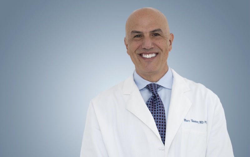
Board-certified ophthalmologist; Adjunct Associate Professor of Ophthalmology, Keck School of Medicine of USC; active California Qualified Medical Evaluator. Full biography · Curriculum vitae
Published August 2, 2026 · Last substantive review August 2, 2026 · Medical reviewer: Marc H. Shomer, MD, PhD, QME
Request a Conflict Check and Case Review
Submit the matter for conflict screening. Please do not send medical records or protected health information until conflicts are cleared and secure transfer instructions are provided.