Understanding Visual Field Testing in Litigation
Visual field testing (perimetry) maps how far peripheral vision extends and how sensitive it is across that expanse: function that acuity testing, which measures only the central point, cannot capture. Field loss can be disabling even when acuity is 20/20, and field data figure prominently in glaucoma, stroke, optic nerve injury, and disability claims. But perimetry has a property attorneys must understand: it depends on the examinee's effort and responses, which is why every field test comes with built-in reliability measures and why fields should always be checked against objective structural imaging.
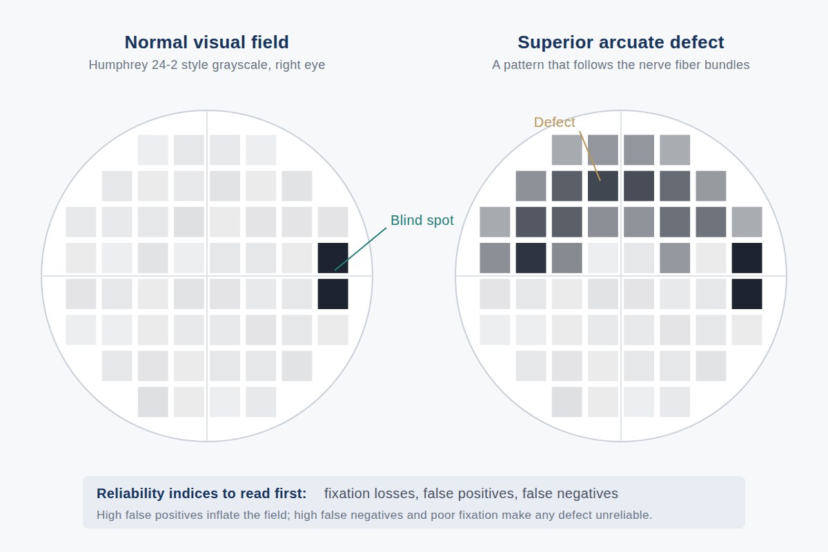
What Perimetry Measures
In automated perimetry, the Humphrey Field Analyzer is the standard instrument, and the 24-2 pattern the most common protocol, the examinee fixates on a central target while lights of varying brightness appear at fixed points across the field. Each button press records that a stimulus was seen. The machine assembles a sensitivity map, compares it to age-matched normal values, and prints deviation plots highlighting where sensitivity falls below expected levels. The result quantifies peripheral function point by point, something no other routine clinical test does.
Reliability Indices: The First Thing to Read
Because the test depends on subjective responses, the machine builds in checks, printed on every result:
- Fixation losses, how often the examinee responded to a stimulus placed in the physiologic blind spot, indicating wandering gaze.
- False positives, button presses when no stimulus was shown ("trigger-happy" responding, which fabricates sensitivity).
- False negatives, failures to respond to bright stimuli in areas already shown to see well, suggesting fatigue or inattention.
High error rates flag an unreliable test. A field with 40 percent false positives is not evidence of anything, however dramatic its printout looks. Reading the reliability indices before the grayscale map is the habit that separates specialist interpretation from a glance at the picture.
Common Patterns and What They Suggest
Field defects have anatomy. An arcuate defect curving around central vision is characteristic of glaucoma and other optic nerve disease. Loss respecting the vertical midline, the same half of the field in both eyes, points to pathology behind the optic chiasm, such as stroke or tumor, a signature of neuro-ophthalmic disease. An altitudinal defect (upper or lower half) suggests ischemic optic neuropathy. Generalized constriction, a field squeezed inward from all sides, appears in advanced disease, but a very constricted "tunnel" field that fails to expand when tested at greater distance, or in a person navigating a room without difficulty, raises the question of non-organic loss. Pattern recognition is where specialist review earns its place: the shape of the defect must fit the alleged cause.
Effort Dependence: The Central Caveat
Perimetry is a psychophysical test, it measures responses, not anatomy. An examinee who does not press the button appears to have field loss whether or not any exists. This does not make fields worthless; it makes them evidence that requires corroboration. Repeat testing should show consistent defects in consistent locations. Defect shape should match a plausible disease. And structure should explain function, which is where OCT comes in.
Corroborating Fields with Structure
Optical coherence tomography (OCT) objectively measures the retinal nerve fiber layer, the axons that carry visual information from retina to brain. Genuine field loss from optic nerve disease is accompanied by corresponding, anatomically appropriate nerve fiber thinning; a devastated field over a robustly normal OCT is a mismatch demanding explanation. Structure-function correlation is the single most powerful tool for validating or questioning field results, and it is discussed in more depth in OCT as Evidence and Evaluating Claimed Vision Loss.
Goldmann Fields and Disability Rating
Goldmann perimetry is a manual technique in which a trained examiner moves stimuli inward from the far periphery, mapping the boundaries of the field. Because it covers the full peripheral extent and permits examiner interaction, Goldmann (or an equivalent kinetic protocol) remains the reference method for disability evaluation, and the AMA Guides' visual field impairment calculations are built on field extent along specified meridians. Automated 24-2 testing, which samples only the central 24 degrees, cannot substitute for full-field data when the question is how much of the total field survives. In whole person impairment ratings for vision, field loss and acuity loss are combined into a single figure.
Key point: A visual field is evidence only to the extent it is reliable, reproducible, anatomically sensible, and corroborated by structure. All four can be assessed from the records: provided the native field printouts, not degraded copies, are produced.
What Attorneys Should Provide
Produce every field study ever performed, not only the most recent, in native or first-generation form, together with OCT data and the clinical notes surrounding each test. Serial fields matter: progression, stability, or inconsistency over time is often the heart of the opinion.
Frequently Asked Questions
What is a visual field test?
A visual field test, or perimetry, maps the extent and sensitivity of peripheral vision by recording an examinee's responses to light stimuli presented across the field of view. Automated Humphrey perimetry is the clinical standard; manual Goldmann perimetry maps the full peripheral extent and remains important in disability evaluation.
Can a visual field test be faked?
Because perimetry records voluntary responses, poor effort or deliberate non-response can produce apparent field loss. The test's built-in reliability indices, reproducibility across sessions, the anatomic plausibility of the defect pattern, and correlation with objective OCT imaging together allow an examiner to judge whether a field reflects genuine loss.
What are reliability indices on a Humphrey field?
Fixation losses, false positives, and false negatives: automated checks printed on every result that measure whether the examinee maintained gaze, pressed the button without stimuli, or missed stimuli in seeing areas. High error rates mean the field cannot be taken at face value, regardless of how the map looks.
Why are Goldmann fields used for disability ratings?
Goldmann perimetry maps the full peripheral extent of the field along specified meridians, which is what the AMA Guides' visual field impairment calculations require. Standard automated 24-2 testing samples only the central 24 degrees and cannot show how much of the total field remains.
Authorities Cited
Primary sources referenced on this page. Links open the official text on the publishing agency’s site.
Educational information only. This page provides general information for attorneys and other medical-legal professionals. It is not medical or legal advice, does not address any particular case, and does not create a physician-patient, attorney-client, or expert-client relationship. Opinions in any matter are formed only after review of the specific records, examination findings, and applicable literature. Past engagements do not guarantee any result.

Board-certified ophthalmologist; Adjunct Associate Professor of Ophthalmology, Keck School of Medicine of USC; active California Qualified Medical Evaluator. Full biography · Curriculum vitae
Published August 2, 2026 · Last substantive review August 2, 2026 · Medical reviewer: Marc H. Shomer, MD, PhD, QME
Request a Conflict Check and Case Review
Submit the matter for conflict screening. Please do not send medical records or protected health information until conflicts are cleared and secure transfer instructions are provided.