The Eye Is the Most Measurable Sensory Organ, and That Protects Both Sides
No other sensory organ can be measured as objectively as the eye. Its structures can be photographed and scanned at micron resolution, its function mapped point by point, and its electrical responses recorded without any voluntary participation by the examinee. For litigation, this has a symmetrical consequence that both sides should appreciate: objective ophthalmic testing exposes unreliable claims, and it vindicates genuine ones that might otherwise be denied as unprovable.
Why the Eye Is Different
Consider the evidentiary problem in most sensory and symptom-based claims. Chronic pain cannot be imaged. Tinnitus cannot be photographed. Diminished smell, cognitive fog, altered sensation, these rest almost entirely on the claimant's report, leaving factfinders to weigh credibility with little independent evidence. The eye is the great exception. Its transparent optics allow direct observation of living tissue, the retina and optic nerve are the only parts of the central nervous system visible from outside the body, and a mature toolkit measures both its structure and its function with instruments that record data, not impressions.
The Structural Instruments
Optical coherence tomography (OCT) produces cross-sectional images of the retina at near-microscopic resolution and quantifies the thickness of its layers in microns, including the retinal nerve fiber layer that reflects optic nerve health. It is objective in the strict sense: the examinee's effort, mood, and motives cannot alter what the scan shows. It is also ubiquitous, routinely performed in clinical care, so contemporaneous scans often exist from before and after the events in dispute. Its evidentiary uses and technical limits are treated fully in OCT as Evidence.
Fundus photography and angiography create a dated visual archive of the retina and optic nerve. A photograph of a healthy macula two years before an incident, set beside a photograph of a scarred one after, is evidence a lay factfinder can literally see. Serial photography documents progression or stability in a form no narrative report matches.
Anterior segment examination leaves its own objective trail: angle recession from old blunt trauma, surgical wounds, corneal scars, findings recorded at the slit lamp that mark past events on the tissue itself, often decisively in ocular trauma disputes.
The Functional Instruments
Automated perimetry maps visual field sensitivity point by point. It requires the examinee's responses, so it is not purely objective: but it audits itself with reliability indices (fixation losses, false positives, false negatives) and reveals whether a claimed defect reproduces across sessions and matches the anatomy of a real disease, as explained in Understanding Visual Field Testing.
Pupillary testing is functional yet involuntary: the pupil's light reflex cannot be faked or suppressed. A relative afferent pupillary defect objectively marks asymmetric optic nerve or retinal dysfunction; its absence is difficult to reconcile with claimed profound loss in one eye.
Electrophysiology, the electroretinogram (ERG) recording the retina's electrical response, and the visual evoked potential (VEP) recording signal arrival at the brain's visual cortex, measures the visual pathway's function with no voluntary response at all. These studies answer the hardest cases: an eye claimed blind that conducts normally, or, just as important, an eye that looks structurally unremarkable but demonstrably does not conduct.
How Objectivity Protects Defendants and Insurers
The defense application is intuitive. Claimed loss that exceeds what structure and involuntary function support is identifiable: the normal OCT beneath a claim of severe central loss, the full-field electroretinogram in an eye reported dark, the visual field whose reliability indices disqualify it. Objective testing converts "we doubt the claim", a credibility contest, into "the measurements do not support the claim," a documented finding. It also quantifies pre-existing disease, supporting fair apportionment rather than payment for damage an incident did not cause.
How Objectivity Protects Plaintiffs and Applicants
The mirror image deserves equal emphasis. Genuine visual injury is sometimes denied precisely because vision is assumed to be subjective, an adjuster discounts what cannot be "proven." Objective testing is the answer to that denial. The claimant with a documented macular scar, corresponding OCT disruption, and a reproducible field defect is not asking anyone to take their word for it; the tissue testifies. Objective findings also protect claimants whose injuries are real but invisible on casual examination, subtle traumatic optic neuropathy, early retinal pathology, where specialist-ordered testing converts an easily dismissed complaint into a demonstrated injury. An honest measurement regime has no side; it disciplines both.
The Honest Limits
Objectivity is not omniscience, and overselling it damages credibility. Structure does not equal function: OCT cannot measure what a person experiences, some genuine pathology hides from every scanner, and measurable damage does not automatically mean disabling loss. Tests have artifacts and error rates; interpretation requires accounting for them. And the objective toolkit can show that reported loss exceeds measurable findings without ever revealing why, motive is outside the instruments' reach, as discussed in Evaluating Claimed Vision Loss. The expert's role is to state what the measurements establish, what they exclude, and what they leave open.
The Practical Upshot for Counsel
Whichever side retains you to an eye case, the objective record is likely to be the decisive evidence, and it either exists in native form or it does not. Preserve imaging early, obtain it completely, and put it in front of a specialist who reads the raw data rather than the summary pages. In a field this measurable, the side that actually measures, carefully, completely, and honestly, argues from evidence while the other side argues from impressions.
Frequently Asked Questions
Why is the eye considered the most objectively measurable sensory organ?
Because its transparent optics allow direct imaging of living tissue: OCT scans quantify retinal layers in microns, photographs archive the retina's appearance over time, and electrophysiology records the visual pathway's electrical responses without any voluntary participation. No comparable toolkit exists for pain, hearing complaints like tinnitus, or other subjective symptoms.
How does objective testing help the defense?
It converts credibility disputes into measurement questions. Claims exceeding what OCT, pupillary testing, and electrophysiology support are documented as unsupported rather than merely doubted, unreliable visual fields are identified by their own indices, and pre-existing disease is quantified for fair apportionment.
How does objective testing help plaintiffs?
It proves injuries that would otherwise be dismissed as unverifiable. Documented structural damage with corresponding functional loss means the claimant is not relying on self-report, and specialist-ordered testing can demonstrate real but subtle injuries, such as traumatic optic neuropathy, that casual examination misses and adjusters deny.
Can objective tests determine whether someone is exaggerating on purpose?
No. Objective testing can establish that reported vision loss exceeds the visual system's measurable capacity, but it cannot reveal motive: the discrepancy may reflect unconscious amplification, psychological illness, or deliberate feigning. A careful evaluator documents the findings and the discrepancy without speculating about intent.
Educational information only. This page provides general information for attorneys and other medical-legal professionals. It is not medical or legal advice, does not address any particular case, and does not create a physician-patient, attorney-client, or expert-client relationship. Opinions in any matter are formed only after review of the specific records, examination findings, and applicable literature. Past engagements do not guarantee any result.
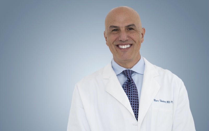
Board-certified ophthalmologist; Adjunct Associate Professor of Ophthalmology, Keck School of Medicine of USC; active California Qualified Medical Evaluator. Full biography · Curriculum vitae
Published August 2, 2026 · Last substantive review August 2, 2026 · Medical reviewer: Marc H. Shomer, MD, PhD, QME
Request a Conflict Check and Case Review
Submit the matter for conflict screening. Please do not send medical records or protected health information until conflicts are cleared and secure transfer instructions are provided.