Macula-On vs Macula-Off: The Distinction That Drives Retinal Detachment Cases
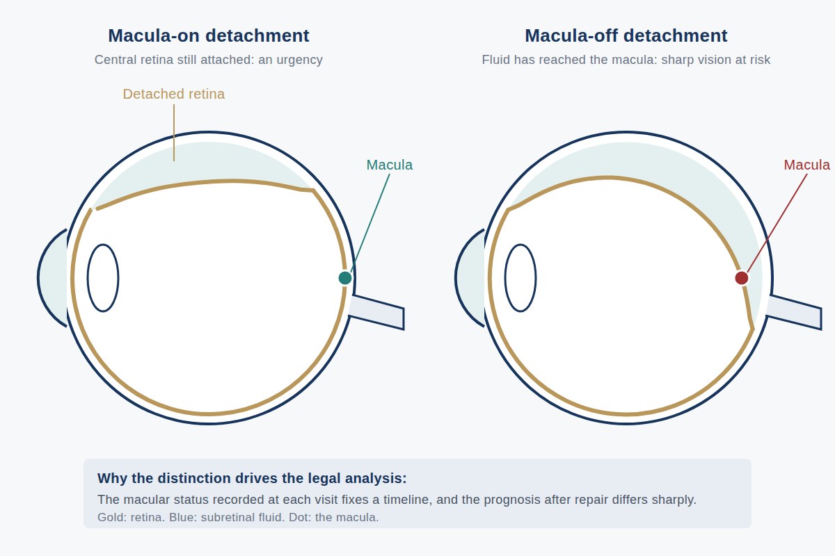
In retinal detachment litigation, one anatomic fact organizes almost everything else: whether the macula was still attached when the patient was seen. A macula-on detachment threatens central vision that is still intact and is treated as a surgical urgency; a macula-off detachment has already taken that vision, and the achievable outcome is different in kind. Because the transition from one state to the other can occur within days, the interval between symptom onset, presentation, diagnosis, and repair becomes the factual spine of any delay claim, and the medical record either documents the macular status at each point or leaves the question genuinely open.
The Anatomy in Plain Terms
The retina is a thin, light-sensitive layer lining the inside of the eye, functioning something like a camera sensor, but it is not uniform. A small central region, the macula, with a still smaller depression at its center called the fovea, holds the dense concentration of photoreceptors responsible for sharp central vision: reading, faces, fine detail, and the acuity measured on a chart. The vast remaining peripheral retina supplies the surrounding field at much lower resolution. That is why a person can lose a large area of peripheral retina and still read 20/20, and why damage confined to a few millimeters of macula can reduce acuity profoundly.
In a rhegmatogenous retinal detachment, the type following a retinal tear or hole, fluid from the vitreous cavity passes through the break and separates the retina from the underlying tissue that nourishes it, cutting the photoreceptors off from their supply. Detachments typically begin peripherally and extend, announced by flashes of light, a shower of new floaters, and then a shadow or curtain advancing across the field. So long as the detachment has not reached the macula, central acuity can remain normal even with a large area of peripheral retina detached. That is the macula-on state.
Why Macula-On Is Treated as an Urgency
In a macula-on detachment, good central vision still exists and is at risk. Once detachment extends under the fovea, photoreceptor damage begins, and while reattachment can restore a great deal, restoration to the prior level is uncertain and becomes less likely as the detached interval lengthens. Ophthalmologists therefore treat macula-on detachments as urgent, arranging prompt repair and often instructing the patient in positioning meant to keep fluid away from the macula in the interim.
A macula-off detachment carries different logic: central vision has already been affected, so the acute pressure to prevent that specific loss has passed and surgery is scheduled promptly but with less compression. That difference is exactly what makes the macula-on case attractive to plaintiffs in delay claims, the theory being that timely diagnosis and repair would have preserved central vision a delay allowed to be lost. Whether the theory holds in a given case depends on evidence rather than on its coherence.
How Macular Status Is Documented
Three sources establish the status, and they are not equally reliable.
- Dilated fundus examination. The ophthalmologist records whether the detachment involves or spares the macula, often with a hand-drawn diagram showing the extent of detachment, the location of breaks, and the relationship of the detachment border to the fovea. A careful drawing is genuinely informative; a note reading only "retinal detachment" is not.
- Optical coherence tomography (OCT). Cross-sectional imaging shows directly whether subretinal fluid extends beneath the fovea, the most definitive documentation available. Its limitation is that OCT is not always obtainable in an eye with a large detachment or in an emergency setting, and the scan must include the fovea to answer the question.
- Fundus photography, including wide-field imaging. Photographs give a dated, reviewable image another ophthalmologist can interpret independently rather than taking a narrative note at face value.
A fourth, indirect source deserves mention: the recorded acuity itself. Central acuity at or near the documented baseline is hard to reconcile with a detached fovea, and markedly reduced acuity in a detached eye is consistent with macular involvement. Acuity corroborates rather than decides, media opacity and hemorrhage also reduce it, but an acuity that contradicts the narrative characterization of macular status is a discrepancy worth resolving. See OCT as Evidence.
Mapping the Timeline onto the Legal Question
A delay claim is an argument about intervals, and the intervals must be built from the record rather than the complaint. The sequence typically includes: symptom onset, as reported and as documented contemporaneously; first contact with any provider, including telephone triage and urgent care or emergency department visits; the first dilated examination by someone able to diagnose a detachment; the date the diagnosis was actually made; referral to a vitreoretinal surgeon; and the repair itself. Each transition is a potential locus of delay attributable to a different actor: the patient, a triage system, a primary provider, a referring ophthalmologist, or surgical scheduling.
The analysis then asks what the macular status was at each documented point. Macula-on at the first examination followed by macula-off at surgery days later frames the question sharply. Where macular status was never documented at the earlier visit, the plaintiff argues inference and the defense argues the macula may already have been off, a contest neither side wins cleanly, which is why the documentation question so often determines the shape of the case.
It is also worth separating two distinct alleged failures that delay cases blur together: failure to diagnose (a patient with flashes, floaters, and a field defect who was not dilated, or was dilated by someone who missed the detachment) and failure to act with appropriate urgency on a diagnosis that was made. They implicate different standards and different evidence, and are analyzed separately under retinal detachment expert review and standard-of-care analysis.
Why the Prognosis Differs
Anatomic success, getting the retina reattached, is not the same thing as visual success. In a macula-on detachment repaired before the fovea detaches, the central photoreceptors were never separated from their supply, and central acuity commonly remains at or near its prior level. In a macula-off detachment the foveal photoreceptors have been detached for some interval, and recovery is partial and variable: residual deficits in acuity, contrast, image distortion (metamorphopsia), and reading function occur even after anatomically perfect repair.
Two consequences follow for damages. The harm in a macula-off case is often not captured by acuity alone, since distortion and reading difficulty can be functionally significant in an eye with a respectable Snellen number, see visual disability evaluation. And the counterfactual matters: the theory is that timely repair would have preserved the pre-detachment vision, and testing it requires knowing what that vision was, which means baseline records again.
The Honest Limits
Several qualifications should temper any confident narrative in either direction.
- Progression is not uniform. Detachments extend at rates depending on the location and size of the break, the state of the vitreous, posture, activity, and factors not fully predictable. Some macula-on detachments stay stable for a considerable period; others involve the macula within hours.
- Documented status at one visit proves status at that visit only. This cuts both ways: it limits the claim that repair one day earlier would have made a difference, and equally limits the argument that the macula was probably already off.
- The macula-on and macula-off categories are not perfectly binary. Detachments may split the fovea, involve it shallowly, or extend to its edge, and the clinical characterization can reasonably vary between examiners looking at the same eye.
- Duration matters within the macula-off category. A fovea detached briefly is not equivalent to one detached for weeks, and prognosis in the macula-off group is not a single number.
- Outcome is multifactorial. Proliferative vitreoretinopathy (scar tissue that can cause redetachment), detachment height, other ocular disease, and the number of operations required all influence the result independent of timing.
What Attorneys Should Gather
For any detachment case: complete records of every encounter from symptom onset forward, including telephone and portal messages and after-hours triage; retinal drawings and all imaging in native format, particularly any OCT including the fovea; the operative report and postoperative course; pre-detachment records from every provider, including optometry, establishing baseline acuity; and surgical scheduling records explaining the interval between the decision to operate and the operation. Where those documents establish macular status and dated intervals, the medicine becomes tractable. Where they do not, the candid opinion says so, itself a useful answer, and often an early one. See retina expert review and What Records to Provide.
Frequently Asked Questions
What does macula-on versus macula-off mean in a retinal detachment?
The macula is the small central region of the retina responsible for sharp central vision. A macula-on detachment has not yet extended under the macula, so central acuity is typically preserved; a macula-off detachment has separated the macula from its underlying nutritional supply, and central vision is already affected. The distinction determines both surgical urgency and expected visual outcome.
Why does macular status matter so much in delayed-diagnosis claims?
Because the plaintiff's theory in most delay cases is that prompt repair would have preserved central vision that a delay allowed to be lost. That theory requires evidence that the macula was still attached at a point when timely intervention was possible, and that it detached during the disputed interval. Without documentation of macular status at each encounter, the theory rests on inference.
How is macular status documented in the record?
By dilated fundus examination with a retinal drawing showing the detachment border relative to the fovea, by optical coherence tomography demonstrating whether subretinal fluid extends beneath the fovea, and by fundus photography that another ophthalmologist can review independently. Recorded visual acuity corroborates these findings but does not substitute for them.
Does documented macula-on status at one visit prove the macula stayed attached?
No, and this is the most commonly overstated inference in these cases. Detachments extend at rates influenced by break location, vitreous state, posture, and activity, and progression is not reliably predictable. A macula-on finding establishes the status on that date only, a limitation that constrains plaintiff and defense arguments equally.
Educational information only. This page provides general information for attorneys and other medical-legal professionals. It is not medical or legal advice, does not address any particular case, and does not create a physician-patient, attorney-client, or expert-client relationship. Opinions in any matter are formed only after review of the specific records, examination findings, and applicable literature. Past engagements do not guarantee any result.

Board-certified ophthalmologist; Adjunct Associate Professor of Ophthalmology, Keck School of Medicine of USC; active California Qualified Medical Evaluator. Full biography · Curriculum vitae
Published August 2, 2026 · Last substantive review August 2, 2026 · Medical reviewer: Marc H. Shomer, MD, PhD, QME
Request a Conflict Check and Case Review
Submit the matter for conflict screening. Please do not send medical records or protected health information until conflicts are cleared and secure transfer instructions are provided.